
By Dr. Belinda Gregory-Head, Dentist at Dental Implant Partners, San Francisco
Key Takeaways
- Early restorative treatment can stop cracked teeth, deep decay, and tooth wear from progressing into fractures, infection, or tooth loss.
- Recurrent decay around fillings or crowns and bite instability often progress without obvious symptoms until major structural damage occurs.
- Replacing missing teeth promptly helps preserve bone, maintain bite stability, and prevent neighboring teeth from shifting out of position.
In restorative dentistry, the most serious problems rarely begin as emergencies. They often start as subtle structural weakness, early decay, minor wear, or small failures around existing dental work. By the time pain becomes obvious, the underlying damage is usually more advanced and more difficult to treat conservatively. That is why I place such strong emphasis on early diagnosis and timely restorative care.
As a restorative dentist, I evaluate these conditions not only by symptoms, but by their effect on tooth strength, occlusion, pulpal health, and long-term prognosis. In this article, I will discuss eight dental conditions I commonly treat before they worsen, and why prompt intervention often preserves more tooth structure and better treatment options.

1. Early Tooth Fractures and Cracked Teeth
Why small structural cracks should never be underestimated
One of the most common conditions I treat before it escalates is early structural fracture in a tooth, particularly cracked cusps and incomplete coronal cracks. These defects may initially present with symptoms that patients describe as intermittent sensitivity on biting, fleeting discomfort when releasing pressure, or generalized awareness that “something feels off” on one side. In many cases, the crack is not grossly visible to the patient, and sometimes it is not dramatic even on routine visual examination. However, under magnification, transillumination, or selective bite testing, these teeth often reveal clear evidence of structural compromise. From a restorative standpoint, a crack is significant not because it is visible, but because it changes how force is distributed through the tooth.
A cracked tooth behaves differently under load than an intact one. When occlusal forces concentrate along unsupported cuspal inclines or traverse a weakened internal line angle, the tooth can flex microscopically with each chewing cycle. That repeated flexure may propagate the crack deeper into dentin, extend toward the pulp, or create a pathway for bacterial ingress and inflammatory pulpal change. What begins as a reversible sensitivity issue can evolve into irreversible pulpitis, cusp fracture, or even a vertical root fracture if left unmanaged. Once the crack extends subgingivally or into the root, the treatment options become significantly more limited and the prognosis more guarded.
My objective in these cases is to diagnose the structural weakness early and stabilize the tooth before catastrophic failure occurs. Depending on the crack pattern, the remaining tooth structure, and the occlusal demands, treatment may range from a bonded onlay to a full-coverage crown designed to splint the cusps and reduce flexural stress. I also evaluate the bite carefully, because a restoration alone is not enough if the tooth continues receiving destructive lateral or concentrated force. When I intervene at the right stage, I can often preserve pulpal vitality and avoid more invasive treatment. That is why a crack that seems minor to a patient can be highly significant to a restorative dentist.
How restorative treatment protects compromised enamel and dentin
The treatment of cracked teeth is not simply about covering the tooth; it is about controlling biomechanics. A properly designed indirect restoration redistributes occlusal force away from unsupported enamel and weakened cusps, converting a structurally vulnerable tooth into one that can tolerate function more predictably. In my treatment planning, I pay close attention to ferrule, cuspal thickness, internal line angles, and the position of functional contacts. A restoration that is too conservative may fail to capture the weakened portions of the tooth, while one that is unnecessarily aggressive sacrifices valuable structure. Restorative dentistry is most effective when it balances conservation with biomechanical protection.
Material selection also matters substantially in these cases. Bonded ceramics, high-strength indirect composites, and traditional crown materials each have advantages depending on the case design, parafunctional load, and remaining enamel available for adhesion. When I can preserve enamel and use adhesive principles effectively, I often gain excellent reinforcement and marginal control. However, if the tooth has significant structural loss, previous large restorations, or evidence of crack propagation into deeper dentin, full cuspal coverage may be the more durable option. The restoration must be designed not only to look natural, but to resist the very forces that caused the problem in the first place.
Equally important is patient-specific risk management after the restoration is placed. Many cracked teeth occur in patients who clench, grind, chew unevenly, or function with unstable posterior support. If the underlying load pattern is not addressed, even a beautifully executed restoration can be challenged prematurely. I frequently discuss occlusal guards, adjustment of interferences, and the relationship between muscle activity and structural fatigue. Treating the crack without treating the cause is incomplete restorative care. My responsibility is not only to repair the damaged tooth, but to improve the environment in which that tooth must function.
What can happen when cracked teeth are ignored
When early fractures are dismissed, the consequences are often more dramatic than patients expect. The most immediate risk is cusp fracture, particularly in posterior teeth that already contain large restorations or undermined enamel. Once a cusp breaks away, the defect is often deeper, less restorable, and more likely to expose dentin or pulp. A case that might have been managed with a conservative indirect restoration can quickly escalate into root canal therapy, crown lengthening, or extraction if the fracture line extends below the gumline. The difference between timely and delayed treatment can be the difference between preservation and tooth loss.
There is also a biologic consequence to crack progression that is frequently overlooked. Microleakage and pulpal irritation can occur even before a tooth breaks visibly, because microscopic movement along the crack line permits bacterial irritants and fluid shifts within dentinal tubules. Patients may notice thermal sensitivity, spontaneous aching, or pain on release that becomes more frequent over time. Once the pulp becomes irreversibly inflamed, I can no longer limit treatment to structural rehabilitation alone. The case becomes endodontic as well as restorative, which changes the long-term restorative design and overall prognosis.
From a long-term standpoint, delaying treatment on a cracked tooth often reduces the quality of the final result. A tooth restored early can frequently be preserved with better margin placement, stronger remaining walls, and a more favorable pulpal status. A tooth restored late is often rebuilt under more compromised circumstances, with less native structure and greater dependence on complex procedures. In restorative dentistry, the prognosis is strongly influenced by how much healthy biology remains when treatment begins. That is why I strongly encourage evaluation of biting pain, unexplained sensitivity, or localized chewing discomfort before the tooth declares itself in a more destructive way.
2. Recurrent Decay Around Existing Fillings and Crowns
Why old dental work often fails at the margins first
Another condition I treat routinely before it worsens is recurrent decay developing around existing restorations. Patients often assume that once a tooth has been filled or crowned, it is permanently protected from future decay. In reality, restorations do not make teeth immune to disease; they simply replace lost structure. Over time, margins can deteriorate, composite can wear, cement can dissolve, and microscopic gaps can form where plaque biofilm becomes more difficult to control. Even excellent restorations have a life cycle, and the interface between tooth and restoration remains biologically vulnerable.
Recurrent decay tends to form in areas where the restoration margin is rough, open, overcontoured, or located in a zone of persistent plaque retention. I frequently see this beneath aging composite restorations, at the crown margin near the gumline, or adjacent to large posterior fillings where cleansability has declined over the years. The danger is that recurrent caries is often hidden. Externally, the tooth may look relatively stable, while radiographs or clinical exploration reveal soft dentin extending under the existing restoration. By the time a patient notices pain or visible discoloration, the lesion may already be extensive.
From a restorative perspective, early intervention is essential because recurrent decay can undermine large portions of remaining tooth structure before it becomes obvious. A small marginal defect today may require straightforward replacement of a filling or a more refined indirect restoration. If postponed, the same defect can progress until the tooth needs endodontic treatment, crown build-up, or extraction due to insufficient restorable structure. In my practice, I do not evaluate an old restoration solely by whether it is still present. I evaluate whether it is still sealing effectively, functioning harmoniously, and preserving the surrounding tooth.
How I assess whether a restoration can be repaired or must be replaced
The management of recurrent decay requires more nuance than simply replacing everything that looks old. A restorative dentist must determine whether the defect is localized and repairable or whether the failure reflects a broader compromise in the restoration’s design, seal, or structural support. I assess the restoration margins, occlusal anatomy, contact relationships, plaque retention pattern, radiographic findings, and the extent of sound residual tooth structure. A conservative repair can be highly effective in selected cases, especially when the defect is limited and the remainder of the restoration is functioning well. However, when the decay undermines critical portions of the restoration or the restoration itself is poorly designed, complete replacement is usually the more predictable choice.
I also consider the material behavior of the original restoration. Composite restorations may exhibit marginal staining that is purely superficial, but they may also show true marginal breakdown, wear, bulk fracture, or interfacial leakage. Full-coverage crowns can appear intact clinically while concealing submarginal decay at the crown-tooth junction. In these cases, treatment planning must account for how much decay is present and whether the remaining tooth will support another direct restoration, an onlay, or a new crown. This is where restorative dentistry becomes strategic rather than reactive. The goal is not just to remove decay, but to place the tooth into a more durable restorative category for the future.
An important part of this process is identifying why the restoration failed. If a restoration is replaced without addressing xerostomia, diet, home care limitations, occlusal overload, or poor contact design, the new restoration may fail for the same reasons. I spend considerable time analyzing risk factors because recurrent decay is rarely just a material problem. It is usually the result of biology interacting with design. That is why thoughtful diagnosis is as important as technical execution when managing failing restorations.
Why delaying treatment can lead to more extensive tooth loss
If recurrent decay is not treated early, it typically expands beneath the restoration where it is shielded from routine cleaning and difficult for the patient to monitor. This hidden progression is what makes the condition particularly deceptive. By the time the old filling fractures, the crown loosens, or symptoms develop, the lesion may have already consumed much of the coronal dentin. What looked like “just replacing an old filling” can suddenly become a crown, post and core, root canal, or extraction. This escalation is common, and it is one of the strongest arguments for periodic restorative evaluation.
There is also a mechanical consequence to recurrent decay that patients do not always appreciate. As caries progresses under a restoration, it reduces the support of the restoration itself and weakens the surrounding tooth walls. The restoration may begin to flex, fracture, or leak more severely, accelerating the cycle of destruction. In posterior teeth, this frequently leads to cusp fracture or loss of proximal wall integrity, which compromises contact and food impaction as well. Once these structural relationships fail, treatment becomes more complex and less conservative.
From my standpoint, the benefit of early intervention is not merely eliminating decay, but preserving architecture. When I can treat recurrent decay while enamel margins are still manageable and the tooth remains structurally coherent, the restorative outcome is stronger and more biologically respectful. When treatment is delayed until the restoration fails catastrophically, I am often rebuilding under compromised conditions. In restorative dentistry, every millimeter of preserved tooth structure matters. That is why recurrent decay should never be dismissed simply because an old filling or crown appears to be “still there.”
3. Moderate to Deep Dental Caries Before Pulpal Involvement
Why does cavity progression become much more serious once it reaches dentin
Dental caries is often spoken about in simplistic terms, but from a restorative perspective, the difference between superficial and advanced decay is profound. Once decay breaches enamel and enters dentin, the lesion progresses more rapidly because dentin is less mineralized and more permeable. The anatomy of dentinal tubules also means that bacterial byproducts and inflammatory signals can move toward the pulp long before a patient feels severe pain. What patients may call a “small cavity” can already represent a biologically active lesion with significant restorative implications. This is especially true in interproximal lesions, where the extent of decay is often underestimated until the tooth is opened.
In moderate to deep caries, the concern is no longer limited to removing infected tissue. I must also consider remaining dentin thickness, pulpal response, cavity design, cuspal support, and the long-term prognosis of the tooth under load. A deep lesion in a small tooth with thin remaining walls is fundamentally different from a similarly deep lesion in a structurally robust tooth. The restorative plan must reflect that difference. In some cases, selective caries removal and pulp-protective protocols are appropriate to preserve vitality. In others, the amount of structural compromise means the tooth needs cuspal protection rather than a routine filling.
Patients are often surprised to learn that timely restorative treatment of a deep lesion can prevent root canal therapy. Once bacterial irritation reaches a threshold that the pulp can no longer tolerate, the tooth shifts from a restorative problem to a pulpal disease problem. My goal is to intervene while the pulp remains recoverable and the tooth can still be sealed predictably. Restorative timing matters enormously here. A lesion treated at the right stage often remains a restorative procedure; the same lesion treated too late may require a much more complex sequence of care.
The restorative considerations that go beyond simply “filling a cavity”
When I restore a tooth affected by moderate or deep caries, I am not simply placing material into an empty space. I am reconstructing lost anatomy in a way that must re-establish marginal integrity, proximal contact, occlusal function, and resistance to fracture. The depth and location of the lesion influence whether a direct restoration is appropriate or whether indirect treatment will provide better longevity. Posterior teeth with extensive intracoronal decay often experience increased cuspal deflection after preparation, especially when one or more marginal ridges are compromised. In those cases, a seemingly routine filling may not be the most biomechanically responsible choice.
Adhesive dentistry has improved our ability to treat these teeth conservatively, but it has not eliminated the need for proper case selection. Bond strength is only one part of the equation. Moisture control, enamel availability, matrix adaptation, cervical margin position, and postoperative load all affect clinical success. In deeper lesions, I also assess whether the restoration will be placed on a substrate of primarily enamel, sound dentin, affected dentin, or a combination of these. Each substrate behaves differently, and long-term seal quality depends on understanding that distinction.
Equally important is preserving tooth vitality while delivering a restoration that can withstand function. In some deep lesions, I use liners or bioactive materials selectively, but these are adjuncts, not substitutes for sound restorative design. The real determinant of success is whether I can eliminate active disease and create a durable coronal seal. A restoration that looks acceptable but leaks microscopically or leaves unsupported tooth structure behind is not a successful treatment in my view. The best restorative outcome is one that stabilizes biology, restores mechanics, and minimizes the chance of re-entry in the near future.
Why untreated decay can quickly become a restorative and endodontic problem
The progression from dentinal caries to pulpal involvement can occur gradually or with surprising speed, depending on the patient’s caries risk, diet, salivary status, and lesion activity. Once the pulp becomes irreversibly inflamed or necrotic, the treatment sequence changes substantially. Now the tooth may require endodontic therapy, core build-up, and cuspal coverage, all while functioning with less native structure than it had originally. That added complexity is often preventable. Treating decay before pulpal breakdown gives me more conservative and more predictable restorative options.
There is also a structural consequence to delaying treatment of deeper caries. As dentin continues to demineralize beneath the enamel shell, the remaining tooth becomes progressively less able to resist functional stress. I often see teeth that have become dangerously undermined without causing dramatic symptoms until a wall fractures during routine chewing. At that point, the lesion is no longer just about decay removal. I am now dealing with collapse of the coronal architecture, subgingival extension, and reduced ability to isolate or bond effectively. The restorative burden becomes significantly greater than it would have been weeks or months earlier.
From a clinical standpoint, early treatment of moderate to deep decay is one of the clearest examples of how restorative dentistry can prevent escalation. A well-timed restoration preserves vitality, structure, and future options. A delayed restoration often becomes a more invasive rehabilitation with a less favorable prognosis and greater financial and biological cost. That is why I encourage patients not to postpone care simply because discomfort is mild or intermittent. The absence of severe pain does not mean the disease process is stable.

4. Tooth Wear, Erosion, and Functional Breakdown
Why worn teeth are more than an aesthetic issue
Tooth wear is one of the most misunderstood conditions I treat. Patients commonly believe that flattened edges, thinning enamel, or shorter teeth are simply signs of aging. In reality, pathologic wear reflects an active imbalance involving attrition, erosion, abrasion, or a combination of these mechanisms. When wear progresses beyond physiologic norms, it can alter occlusal stability, reduce enamel thickness, expose dentin, and compromise the vertical and horizontal relationships that support efficient function. Restorative dentists evaluate wear not just by appearance, but by the rate of progression, the pattern of loss, and its effect on the entire stomatognathic system.
Attrition from clenching and grinding creates a very different restorative problem from acid-mediated erosion, even if both ultimately produce shortened or damaged teeth. Attrition tends to generate flattened contact surfaces, fracture lines, and concentrated stress, particularly in patients with parafunctional loading. Erosion, by contrast, softens enamel chemically and may produce cupping, thinning, and loss of surface definition, often in patients with dietary acid exposure or reflux. Many patients present with a mixed picture, where softened teeth are then mechanically worn more rapidly. That combination can accelerate the breakdown significantly and increase restorative complexity if not addressed early.
I approach tooth wear as a condition that affects both structure and biomechanics. As enamel is lost, dentin becomes more exposed and the tooth becomes more susceptible to sensitivity, further wear, and fracture. The bite may also shift subtly as posterior support changes or anterior guidance deteriorates. This can create a feedback loop in which wear worsens function and unstable function worsens wear. Early restorative intervention allows me to stabilize these relationships before the patient develops severe occlusal collapse, widespread restorative need, or significant loss of restorative space.
How restorative dentistry can rebuild strength and occlusal stability
The restorative management of tooth wear requires much more than adding material where structure is missing. I must first determine the etiology, assess the existing vertical dimension, evaluate centric stops and excursive pathways, and decide how much rehabilitation is necessary to restore function without over-treating. In localized wear cases, conservative additive techniques such as bonded composite or selective indirect restorations can often restore anatomy effectively. In more advanced cases, multiple teeth may require coordinated restoration to re-establish stable occlusal relationships. The design must be comprehensive, even if the treatment is phased.
One of the most valuable principles in wear management is preservation through additive dentistry whenever feasible. If I can increase contour and function by bonding to remaining enamel rather than aggressively preparing already shortened teeth, I may preserve biology and reduce treatment burden. However, additive approaches are only appropriate when the occlusal scheme, material thickness, and functional demands support them. In heavily loaded or severely eroded dentitions, indirect restorations may be needed to provide more durable coverage and structural reinforcement. The key is that the restorative plan must follow functional diagnosis, not just aesthetic concern.
Occlusal refinement is essential in these cases. If worn teeth are restored without controlling the contact pattern that caused the wear, the new restorations are likely to fail or the patient will continue to damage adjacent teeth. I evaluate envelope of function, parafunctional patterns, posterior support, and the need for protective appliances after treatment. Restorative success depends on creating a bite that is not only comfortable, but mechanically stable. In my experience, properly treating tooth wear early can prevent a future scenario in which nearly every tooth requires major rehabilitation.
What happens when wear is allowed to progress unchecked
When pathologic wear is ignored, the dentition often transitions from localized damage to generalized breakdown. Incisal edges shorten, posterior cusps flatten, and the enamel reserve that once protected the teeth disappears. As dentin becomes increasingly exposed, sensitivity may worsen and wear can accelerate because dentin abrades more readily than enamel. Patients may also begin to notice chipping, loss of chewing efficiency, or a change in the way their teeth fit together. These are signs that the condition is no longer purely cosmetic or age-related.
At a more advanced stage, wear can complicate restorative care by reducing available restorative space and making it difficult to place durable materials without altering the bite strategically. Severely worn teeth often require careful sequencing, trial equilibration, or staged rehabilitation to recover function in a controlled way. The longer the wear continues, the more likely the case will involve multiple quadrants, compromised aesthetics, and a greater need for interdisciplinary support. That increases both the biological cost and the technical demands of treatment. What might have been managed conservatively early on can become a full-mouth reconstruction problem later.
There is also a quality-of-life dimension to advanced wear that should not be underestimated. Patients may experience muscle fatigue, unstable contacts, food limitation, and increasing anxiety about fracture or visible tooth damage. By the time they seek care, they are often dealing with a condition that has been mechanically active for years. As a restorative dentist, I prefer to intervene when the changes are still limited and the dentition remains adaptable. Early diagnosis and stabilization preserve options, reduce complexity, and allow us to restore function in a far more controlled way.
5. Failing or Fractured Restorations
Why broken fillings and crowns signal a larger structural issue
A fractured filling or damaged crown is rarely an isolated event. In most cases, it is a sign that the restoration, the underlying tooth, the occlusal load, or some combination of those factors has exceeded its functional threshold. Patients often focus on the visible break and assume the solution is merely to replace what fell out or chipped. From my perspective, the more important question is why the failure occurred. Restorations fail for reasons that include material fatigue, recurrent decay, insufficient remaining tooth support, heavy occlusal loading, poor contact design, and undiagnosed parafunction. Identifying the mechanism matters because repeating the same restorative design in the same environment may produce the same failure again.
I see this often in large posterior composites that begin with a small marginal fracture and progress to cusp failure or food impaction. I also see crowns that loosen because the underlying tooth has decayed, because retention form is inadequate, or because the tooth has cracked beneath the restoration. An old restoration may appear functional for years while the supporting tooth structure quietly weakens. When the failure finally becomes obvious, the case may already involve much more than surface damage. That is why a fractured restoration deserves a thorough restorative re-evaluation rather than a simple patch in every case.
Restorative dentistry is fundamentally about creating durable interfaces between biomaterial and biologic tissue. Once that interface has failed, I need to determine whether it can be repaired conservatively, redesigned more effectively, or whether the tooth has entered a more compromised restorative category. Timing matters greatly. If I evaluate the problem when failure is still localized, the options are broader and the tooth structure is usually better preserved. If the patient waits until the restoration breaks extensively or the tooth becomes symptomatic, treatment can escalate quickly.
How I determine the best replacement strategy
When managing a failing restoration, I first assess the remaining tooth, not the restoration alone. The most important questions are how much sound structure remains, where the margins can be placed, whether the tooth is vital and crack-free, and how the tooth functions under load. A replacement filling may be completely appropriate in a conservative case, but in a tooth with wide isthmus width, loss of marginal ridge support, or prior fracture history, a bonded onlay or crown may offer a more predictable long-term result. The replacement strategy must fit the current structural reality of the tooth, not the historical treatment it once received.
I also consider the patient’s bite pattern and risk profile. A restoration placed in a patient with significant bruxism, posterior instability, or a history of repeated restoration failure must be designed differently from one placed in a low-load environment. Material selection, thickness, cusp coverage, contact placement, and surface morphology all become more consequential in these patients. In other words, restorative design is not generic. The success of the new restoration depends on matching the material and preparation design to the biologic and functional demands of the case.
Repair versus replacement is another important decision point. In certain circumstances, a conservative repair can preserve healthy restorative material and avoid unnecessary removal of tooth structure. However, repair only works when the failure is limited and the remaining restoration is reliable. If the restoration is undermined, leaking, poorly contoured, or structurally fatigued, replacement is usually the better choice. My goal is always to preserve tooth structure intelligently, not sentimentally. Preserving a failing restoration at the expense of the tooth is never true conservatism.
Why postponing repair often leads to more invasive treatment
Patients sometimes delay treatment of a broken filling or chipped crown because the tooth is not painful. Unfortunately, symptoms are a poor indicator of how vulnerable the tooth may be. Once a restoration loses integrity, the underlying tooth is more exposed to bacterial infiltration, concentrated occlusal stress, and further fracture. A small fracture line at the margin can evolve into recurrent decay, cusp loss, or pulpal irritation. In many cases, the most serious damage occurs after the restoration has already shown the first sign of failure.
Open margins and fractured restorative surfaces also change the local biofilm environment. Rough areas retain plaque more readily, food becomes impacted, and the gingival tissues may become inflamed around a restoration that no longer supports a healthy emergence profile or contact. This not only threatens the tooth itself, but can compromise the surrounding periodontal architecture that restorative success depends upon. Once tissue inflammation or subgingival extension is present, treatment becomes more difficult technically and biologically. That added complexity is often avoidable when the failure is addressed promptly.
From a long-range restorative standpoint, early management of a failing restoration helps preserve the tooth in a stronger restorative category. A tooth that needs a well-designed replacement today may need a root canal, post, crown lengthening, or extraction later if the failure is ignored. I explain this to patients frequently: the goal is not to replace dentistry unnecessarily, but to intervene when preservation is still realistic. Restorations do not fail all at once. They usually fail progressively, and that progression is precisely where restorative dentistry can make the greatest difference.
6. Missing Teeth and Early Post-Extraction Changes
Why a single missing tooth affects more than the empty space
Many patients view a missing tooth as a localized issue, especially when it is not in a highly visible part of the mouth. From a restorative standpoint, however, a missing tooth initiates a chain of biologic and functional changes that can affect the entire arch. Adjacent teeth may drift, opposing teeth may supra-erupt, occlusal contacts may redistribute, and the edentulous ridge begins to remodel almost immediately. These changes do not always produce symptoms at first, but they can complicate future treatment and compromise long-term function if ignored. Replacing a missing tooth early is often easier and more conservative than correcting the secondary problems that develop later.
The loss of a tooth also alters how chewing forces are shared across the dentition. When one posterior tooth is missing, nearby teeth may assume greater functional load than they were designed to bear, particularly if the patient already has a history of wear, clenching, or restorative compromise. Over time, this may contribute to fracture, tooth migration, periodontal stress, or additional restorative failure in adjacent areas. In the anterior region, a missing tooth may affect phonetics, incisal guidance, and esthetics in ways that subtly destabilize the bite. Tooth loss is therefore not simply a gap problem; it is a system problem.
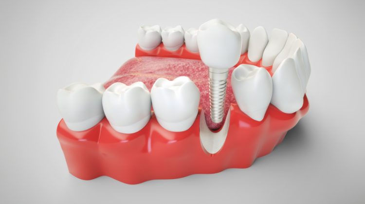
As a restorative dentist, I am especially attentive to what happens in the months after a tooth is lost. Bone resorption, soft-tissue collapse, altered contact relationships, and changing occlusal patterns all influence the eventual restorative options. A site that could have supported a straightforward implant restoration early may require grafting or more complex prosthetic planning later if it is neglected. Likewise, an otherwise simple fixed restoration may become less favorable if adjacent teeth drift or rotate into the space. Timing matters because missing teeth tend to create a moving target over time.
How restorative planning preserves function and arch stability
The treatment of a missing tooth begins with diagnosis, not replacement selection. I evaluate the site, the quality and quantity of remaining bone, the condition of adjacent teeth, the occlusal demands, smile line considerations, and whether the patient would benefit most from an implant-supported restoration, tooth-supported prosthesis, or another approach. In contemporary restorative dentistry, preserving adjacent teeth is a major priority, which is one reason implant therapy has become so valuable when the clinical conditions are favorable. However, the best replacement option depends on the biology and mechanics of the specific case, not a one-size-fits-all philosophy.
What matters most is restoring stable function while minimizing compromise elsewhere in the dentition. If I can replace the missing tooth in a way that preserves the bone, maintains arch form, and restores proper load distribution, I improve not only the appearance of the smile but also the prognosis of the surrounding teeth. Implant-supported restorations can be particularly advantageous because they do not require reduction of neighboring teeth and help support the alveolar architecture. In other situations, a carefully designed bridge or removable prosthetic solution may be indicated depending on the patient’s anatomy, systemic health, and restorative priorities.
I also think ahead when planning these cases. The replacement should integrate with the patient’s occlusion, oral hygiene capabilities, and long-term maintenance needs. An implant crown that is difficult to clean, a pontic design that traps plaque, or a replacement that creates awkward occlusal contacts may solve one problem while introducing another. Restorative dentistry should improve the mouth as a whole. Replacing a tooth successfully means restoring stability, not just occupying space.
Why waiting too long after tooth loss narrows the treatment options
The most immediate consequence of delaying tooth replacement is alveolar bone loss. Once the root is gone, the surrounding bone begins to resorb because it is no longer maintained by physiologic stimulation through the periodontal ligament. This can reduce ridge width and height, complicate implant placement, and require grafting to restore adequate foundation. Soft tissues also lose support, which can affect emergence profile, esthetics, and the natural appearance of the final restoration. These changes are often progressive and significantly influence the complexity of treatment.
Equally problematic is the way neighboring and opposing teeth respond over time. Teeth adjacent to the space may tip or drift, leading to altered contacts, food impaction, and a less favorable restorative environment. Opposing teeth may supra-erupt into the open space, creating occlusal interference and reducing restorative room. Once these positional changes occur, replacing the missing tooth may require orthodontic correction, occlusal adjustment, or more involved prosthetic design. A treatment that was once relatively straightforward can become multidisciplinary because of delay alone.
From a restorative perspective, replacing a missing tooth early is often an act of prevention as much as rehabilitation. It preserves the architecture of the site, stabilizes the bite, and reduces the likelihood that other teeth will be drawn into the problem. In my practice, I encourage patients not to think of tooth loss as a static condition. The mouth adapts to the absence, and those adaptations are rarely beneficial in the long term. Timely restorative care helps preserve simplicity, predictability, and structural balance.
7. Bite Instability and Occlusal Trauma
Why an imbalanced bite can damage otherwise healthy teeth
Occlusal trauma is one of the most underdiagnosed contributors to restorative breakdown. A patient may present with fractured enamel, abfraction-like cervical defects, mobility, soreness on chewing, or repeated failure of restorations, and yet the root cause is not decay or poor hygiene but an unstable force pattern. Teeth are designed to tolerate compressive forces along their long axis far better than they tolerate off-axis lateral stress, repeated parafunctional loading, or premature contacts that concentrate force disproportionately. When these destructive patterns persist, even structurally sound teeth can begin to fail. In restorative dentistry, ignoring occlusion is one of the fastest ways to compromise otherwise well-executed work.
Bite instability can arise from many sources, including missing teeth, drifting teeth, wear, uneven restorations, orthodontic relapse, parafunction, and changes in joint or muscle function. Patients may not be aware of the problem because the symptoms are often diffuse rather than dramatic. They may report jaw fatigue, sensitivity in multiple teeth, repeated chips, or the feeling that one tooth “hits first.” From my standpoint, those complaints are clinically significant because they suggest that the dentition is functioning under an unfavorable load scheme. Restoring teeth in that environment without correcting the force pattern is often a temporary solution at best.
The reason occlusal trauma matters so much is that it changes the prognosis of both teeth and restorations. A crown on a tooth with unresolved excursive interference may loosen, fracture, or precipitate crack propagation beneath the restoration. A filling in a tooth receiving concentrated stress may debond or fail marginally even if the margins were ideal at placement. In many of these cases, the problem is not the material but the mechanics. Restorative dentists must think in terms of load pathways as well as tooth anatomy.
How restorative dentists identify and manage destructive force patterns
When I suspect occlusal trauma, I do not limit my examination to static contact marks on articulating paper. I evaluate wear facets, muscle tenderness, fremitus, mobility patterns, fracture history, joint symptoms, and the dynamic relationship of the teeth during function. I want to know where the heavy contacts are, but also how the patient moves, how the anterior teeth guide those movements, and whether posterior teeth are being subjected to inappropriate lateral load. In many cases, the clinical signs tell a much richer story than the patient’s symptoms alone. The restorative plan must be informed by that functional diagnosis.
Treatment may involve selective adjustment, occlusal refinement of existing restorations, splint therapy, restorative recontouring, or more comprehensive rehabilitation when the instability is widespread. The exact approach depends on whether the problem is localized or systemic. A single high restoration can create a localized traumatic contact, but generalized wear and bite collapse require a broader functional solution. I often coordinate restorative treatment with protective night guards because nighttime parafunctional forces can be substantial even in patients who are largely asymptomatic. Restorative success depends on respecting those force realities.
The design of restorations in these patients must also be deliberate. Contact locations, cusp inclines, anterior guidance, and the distribution of posterior support all influence how force moves through the dentition. I aim to create restorations that harmonize with a stable occlusal scheme rather than compete with it. This is one reason I do not view restorative dentistry as separate from function. In many cases, the durability of the restoration depends as much on the bite as on the preparation or material.
What happens when bite problems are left untreated
When bite instability is allowed to continue, the resulting damage tends to accumulate across multiple teeth rather than stay confined to one area. Patients may experience repeated chipping of front teeth, recurrent loosening of crowns, widening abfraction defects, increased mobility, or a generalized sense that their bite is changing over time. These are not isolated events. They are often expressions of a load environment that the dentition can no longer accommodate without consequence. Restoring one tooth at a time without correcting that environment usually leads to repetition rather than resolution.
There can also be a periodontal dimension to occlusal trauma, particularly when excessive force acts on teeth with reduced support. While trauma does not initiate periodontal disease, it can exacerbate the functional burden on already compromised teeth and contribute to mobility or discomfort. In restorative treatment planning, this matters because the support system of the tooth and the load applied to it must be in balance. A structurally excellent restoration placed on a tooth with traumatic loading and reduced support may not achieve the longevity either the patient or clinician expects.
Ultimately, untreated bite instability narrows restorative options because it creates ongoing damage faster than dentistry can reasonably keep up with. The patient enters a cycle of repair, fracture, replacement, and further breakdown. I try to interrupt that cycle by identifying occlusal trauma early, before it produces widespread wear, multiple cracked teeth, or repeated restorative failures. From my perspective, one of the most valuable things a restorative dentist can do is recognize when the problem is not the tooth alone, but the functional system acting on it.
8. Root Surface Decay and Cervical Structural Breakdown
Why root caries is often more aggressive than patients realize
Root surface decay is a condition I take very seriously because it can advance quickly and undermine teeth in ways that are less visible and more difficult to restore than coronal decay. When gingival recession exposes root surfaces, the substrate is no longer enamel but cementum and dentin, both of which are more vulnerable to demineralization. In patients with dry mouth, periodontal attachment loss, high cariogenic challenge, or plaque retention around restorations, root caries can become a particularly aggressive disease process. It often progresses laterally and circumferentially, compromising the tooth near the gumline where restorative access and isolation are less ideal.
This area is mechanically important as well. The cervical portion of the tooth experiences stress concentration during function, especially in patients with heavy occlusal load or flexural strain. When root caries, abrasion, erosion, or structural fatigue affects this region, the tooth may develop not only decay but also loss of cervical integrity. Patients may present with sensitivity, food trapping, roughness, or visible notching near the gumline. What they often do not appreciate is that these lesions can deepen quietly and make the tooth harder to restore conservatively over time.
From a restorative standpoint, root surface lesions are challenging because they sit at the interface of caries management, margin control, periodontal health, and long-term cleansability. A restoration in this area must seal effectively, contour naturally, and remain maintainable in a zone where moisture control is often limited. Early treatment gives me a better opportunity to restore the defect while the tooth still has sufficient structure and before the lesion extends subgingivally or around the tooth. Once that happens, the restorative prognosis becomes more complicated.
How I restore cervical lesions while protecting the surrounding tissues
The treatment of root caries and cervical breakdown is highly technique-sensitive. I must remove or control diseased tissue, establish a clean restorative margin, and shape the restoration in a way that supports gingival health rather than inflaming the tissue. Overbulked cervical restorations, open margins, or rough surfaces in this area can make plaque control more difficult and increase inflammation. That is why material handling and contour are so important. The restoration must be biologically compatible, not merely retentive.
Material choice is guided by the specific clinical environment. In some cases, adhesive composite offers excellent polishability and contour control. In others, especially where moisture management is difficult or the lesion extends closer to the root surface, glass ionomer-based materials or layered approaches may provide advantages because of fluoride release and chemical adhesion. However, no material compensates for poor access or poor risk control. If the patient has significant xerostomia, plaque accumulation, or high acid exposure, I address those factors concurrently because otherwise recurrence is more likely.
I also look carefully at the functional context of these lesions. Cervical breakdown may be worsened by occlusal stress, particularly when the tooth is subjected to non-axial loading. In those cases, restorative treatment should be paired with occlusal management to reduce the forces acting on the restored area. A cervical restoration placed into a high-stress environment without load control may debond, fracture, or continue to wear. In my view, successful treatment of cervical lesions depends on integrating material science, tissue management, and biomechanics.
Why untreated root decay can jeopardize tooth retention
If root surface decay is left untreated, it can spread rapidly because the involved substrate is less resistant than enamel. What begins as a shallow, localized lesion can become a broad circumferential defect that compromises the tooth’s structural integrity near the cervical region. As the lesion deepens, it becomes increasingly difficult to isolate, restore, and maintain. The gingival margin may become inflamed, the defect may extend below the tissue, and restorative access may be compromised without periodontal intervention. At that stage, a condition that was once straightforward becomes significantly more difficult to manage conservatively.
The danger is not only recurrent decay but eventual loss of strategic tooth structure in a critical zone. The cervical area contributes substantially to how the crown of the tooth resists functional forces. Once that region is weakened by decay and repeated breakdown, the tooth may become more susceptible to fracture or poor restorative prognosis. In elderly patients or those with multiple recession defects, this can have a cumulative effect across the dentition. Teeth that appear restorable individually may begin to fail collectively if the underlying pattern is ignored.
For that reason, I regard early intervention in root caries as a tooth-preserving measure rather than a minor repair. Addressing the lesion before it becomes circumferential or subgingival preserves restorative simplicity, improves margin control, and reduces the likelihood of tooth loss. Patients sometimes underestimate these defects because they do not resemble the classic “cavity” they expect to see. Clinically, however, root caries can be among the more consequential conditions we treat. Early management is one of the best ways to preserve long-term dentition stability.
To Conclude: Restorative Care Is Most Predictable Before Disease Becomes Advanced
In restorative dentistry, the most favorable outcomes usually occur before a condition becomes dramatic. A crack is easier to stabilize before it splits a cusp. Recurrent decay is easier to manage before it undermines the entire restoration. Wear is more conservative to rebuild before the bite collapses, and a missing tooth is simpler to replace before bone and tooth positions change. This is the pattern I see repeatedly in practice. The earlier I intervene, the more likely I am to preserve natural structure, avoid unnecessary complexity, and provide a restoration that performs well for years.
What patients often call “waiting to see if it gets worse” can be a costly strategy in the mouth because dental conditions frequently progress in hidden ways. Structural failure, pulpal inflammation, subgingival extension, occlusal instability, and ridge resorption often develop long before the problem becomes urgent enough to force treatment. By the time symptoms become obvious, the biologic and mechanical environment has usually become less favorable. Restorative dentistry is not simply about replacing what is broken. It is about recognizing when the tooth, the bite, or the existing dental work is entering a vulnerable stage and acting before options narrow.
As Dr. Belinda Gregory-Head of Dental Implant Partners in San Francisco, I believe the best restorative treatment is often the treatment that prevents a more invasive procedure later. That requires careful diagnosis, an understanding of biomechanics and oral biology, and a willingness to treat problems at the stage where preservation is still possible. Every restoration I place must support not only the tooth in question, but the long-term health and function of the entire dentition. When patients seek care early, restorative dentistry can be remarkably conservative, precise, and durable. That is why identifying and treating these eight conditions before they worsen is not only good clinical practice, but one of the most effective ways to protect long-term oral health.

Why Patients Trust Dental Implant Partners for Restorative Care
At Dental Implant Partners, we understand that treating dental conditions early is one of the best ways to preserve healthy tooth structure, protect long-term function, and prevent more complex problems from developing. For more than 25 years, this practice has been the prosthetic practice of Dr. Belinda Gregory-Head, and today I am proud to work alongside a larger team of experienced prosthodontists and general dentists who share a deep commitment to exceptional patient care. We provide a full range of restorative services, from simple fillings and veneers to comprehensive dental rehabilitations on dental implants. We also have extensive expertise in dentures for patients who are not ideal candidates for implant treatment. Our goal is always to deliver thoughtful, precise care that reflects both technical excellence and an individualized understanding of each patient’s needs.
What makes our practice especially meaningful to me is the consistency and experience of the team our patients see every day. Our hygienists are both trained as dentists and have been with us for many years, which gives our patients an uncommon level of continuity, trust, and clinical insight throughout their care. We value long-term relationships and believe restorative dentistry should be careful, ethical, and built around lasting results rather than quick fixes. Every treatment recommendation we make is guided by what will best support the patient’s oral health, comfort, and quality of life over time. We are honored to provide this care in our beautiful suite overlooking the San Francisco Bay, where we have created an environment that reflects the high level of attention we bring to our dentistry.
We love restoring smiles, and we love the work we do because of the confidence, comfort, and function it can give back to our patients. If you have noticed changes such as tooth sensitivity, cracked dental work, worn teeth, missing teeth, or other signs that something may be worsening, I encourage you not to wait until the problem becomes more complex. Early restorative treatment often gives us more conservative and more predictable options. At Dental Implant Partners, we would love to help you protect your oral health with treatment that is personalized, comprehensive, and designed for long-term success. Contact our office to schedule a consultation and learn how we can help with your dental needs.