By Dr. Belinda Gregory-Head, DDS at Dental Implant Partners, San Francisco
- Bone grafting rebuilds deficient jawbone so dental implants can be positioned for stable function, hygiene, esthetics, and long-term tissue support.
- The appropriate graft material and surgical timing depend on defect anatomy, blood supply, implant stability, medical risks, and restorative goals.
- Predictable bone-grafting outcomes require precise planning, rigid graft protection, controlled healing, complication management, and lifelong implant maintenance.
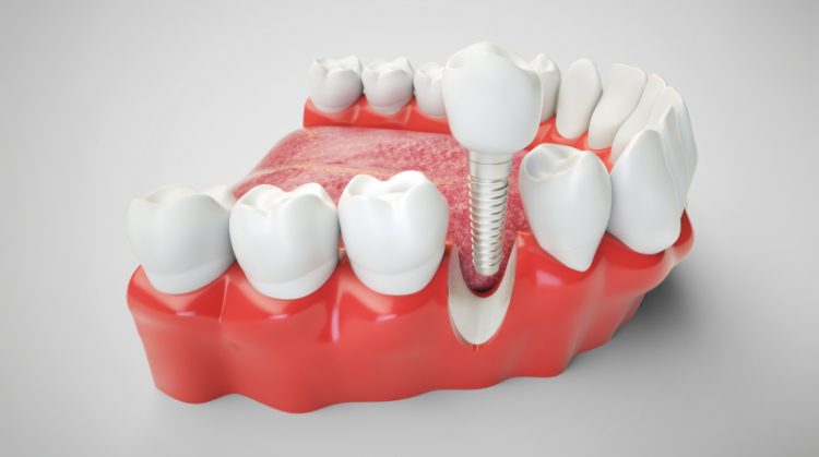
Bone grafting in implant dentistry is not simply a matter of “adding bone” to an area that looks thin on an X-ray. It is a biologically and prosthetically driven procedure intended to create a stable three-dimensional foundation for an implant, its restoration, and the surrounding soft tissues. In my practice, I evaluate the proposed tooth position first and then determine whether the existing ridge can support an implant in that restorative position.
This distinction matters because an implant can sometimes be placed into available bone yet still be poorly positioned for hygiene, esthetics, force distribution, or long-term tissue stability. The purpose of this article is to explain how experienced clinicians analyze these cases, select grafting methods, manage risk, and determine when augmentation is truly justified. It is educational in nature and cannot replace a clinical examination, medical review, and individualized treatment plan.
Why Bone Grafting Is Sometimes Necessary
Implant Placement Must Be Prosthetically Driven
I begin implant planning with the final crown, bridge, or overdenture in mind rather than with the bone alone. The implant must emerge in a position that supports a natural contour, cleansable prosthesis, favorable screw-access trajectory, and controlled occlusal loading. When the residual ridge is too narrow, too short, or positioned outside that restorative envelope, placing the implant wherever bone happens to remain can create a compromised result. Facial placement may produce recession and visible implant components, while excessive palatal or lingual placement can create bulky contours that trap plaque and alter speech. Bone grafting is therefore often a means of correcting the relationship between anatomy and the planned restoration, not merely increasing a radiographic measurement. Contemporary implant literature emphasizes that circumferential bone support and appropriately positioned peri-implant tissues are central to predictable long-term stability.
Tooth Loss Changes Ridge Anatomy
After a tooth is removed, the socket does not heal by perfectly recreating its original dimensions. The facial socket wall, particularly in the anterior maxilla, is often thin and susceptible to remodeling, so horizontal contraction can be clinically significant even when the extraction was atraumatic. Infection, periodontal bone loss, root fracture, trauma, and previous surgery can magnify the defect before healing even begins. In long-standing edentulous areas, resorption may continue until the ridge becomes too narrow for ideal implant positioning or too close to structures such as the maxillary sinus or inferior alveolar nerve. Ridge-preservation procedures can reduce dimensional change, but they do not eliminate remodeling and do not guarantee that later augmentation will be unnecessary. I explain this early because socket preservation is a risk-reduction strategy, not a promise that the site will retain its original anatomy.
Grafting Is Not Automatically the Best Answer
A sophisticated treatment plan also considers whether grafting can be avoided without sacrificing the restorative result. Shorter implants, narrower implants, altered implant distribution, tilted implants in selected full-arch cases, or a different prosthetic design may sometimes reduce surgical morbidity. Those alternatives are not universally preferable, because changing implant dimensions or position can alter biomechanics, emergence profile, cleansability, and the amount of restorative compromise. In the posterior maxilla, for example, evidence suggests that short implants may perform comparably to longer implants placed with sinus augmentation in selected situations, but case selection remains critical. I therefore compare the biological cost of grafting with the mechanical and prosthetic cost of avoiding it. The correct question is not whether grafting is technically possible, but whether it creates a meaningfully better and more maintainable treatment.
How I Determine Whether a Patient Needs a Bone Graft
Clinical Examination and Restorative Analysis
The diagnostic process begins with far more than measuring ridge width. I assess the smile line, gingival display, tissue phenotype, keratinized mucosa, vestibular depth, adjacent tooth position, interarch space, occlusion, parafunctional habits, and the patient’s ability to clean a future restoration. Palpation can reveal undercuts and ridge concavities that are not obvious on two-dimensional radiographs. A diagnostic tooth setup or digital wax-up helps establish the intended crown position before any implant osteotomy is planned. From that position, I evaluate whether the implant can be surrounded by an adequate bone envelope and whether the soft tissue can be shaped without excessive restorative compensation. This is why implant planning should be coordinated between surgical and restorative disciplines rather than divided into isolated procedures.
Three-Dimensional Imaging and Anatomical Mapping
Cone-beam computed tomography is particularly valuable when the anatomy is complex, a graft is being considered, or critical structures could influence implant position. Cross-sectional images allow me to examine ridge width and height, facial concavities, sinus anatomy, septa, nasal-floor position, nerve location, and the morphology of residual defects. I also evaluate bone-density patterns cautiously, because grayscale values from CBCT are not equivalent to standardized medical CT density measurements. Digital planning can superimpose the proposed restoration onto the scan, revealing whether a seemingly adequate ridge actually supports the correct implant trajectory. The scan does not make the treatment decision by itself; it improves the quality of the anatomical question being asked. Professional guidance supports three-dimensional imaging when implant receptor sites or augmentation sites require cross-sectional assessment.
Defect Classification and Risk Stratification
Not all bone defects behave the same way, even when they have similar dimensions. A contained defect with intact walls has a better blood supply and space-maintaining geometry than a non-contained horizontal or vertical deficiency. Horizontal augmentation is generally more predictable than major vertical reconstruction because vertical grafts must resist soft-tissue pressure while revascularizing over a greater distance. I also distinguish between a localized dehiscence around a simultaneously placed implant and a severely atrophic ridge requiring staged reconstruction. Patient-level factors such as smoking, diabetes control, periodontal history, medication exposure, and previous radiation can alter the risk category. The final recommendation reflects defect anatomy, regenerative capacity, surgical complexity, restorative benefit, and the patient’s tolerance for time, cost, and morbidity.
The Biology of Bone-Graft Healing
Osteogenesis, Osteoinduction, and Osteoconduction
Bone-graft materials contribute to healing through different biological mechanisms, and the terms should not be used interchangeably. Osteogenesis refers to living bone-forming cells that can directly contribute to new bone, a property associated most clearly with freshly harvested autogenous bone. Osteoinduction describes signaling that recruits progenitor cells and promotes their differentiation toward a bone-forming lineage. Osteoconduction refers to a scaffold that permits vascular and cellular ingrowth while the patient’s own tissues form new bone around or through it. Many commonly used substitutes are primarily osteoconductive and depend on the recipient bed for vascularity and cellular activity. I select materials according to the biological demands of the defect rather than assuming that one product is universally superior.
Vascularity and Mechanical Stability
A graft succeeds only when it is stabilized, protected, and connected to a viable blood supply. Micromotion can disrupt early vessel formation and favor fibrous tissue rather than organized bone regeneration. This is why fixation screws, tacks, tenting devices, titanium mesh, or carefully adapted membranes may be used in larger defects. The recipient site may also be perforated in selected techniques to encourage bleeding and communication with cancellous bone, although the clinical value of every decortication protocol is not identical. Primary wound closure is critical because exposure can introduce bacteria and collapse the protected space. Experimental sophistication does not overcome basic biology: stable clot formation, vascular access, space maintenance, and soft-tissue protection remain foundational.
Remodeling Does Not Mean Every Graft Particle Disappears
Patients are sometimes told that a graft “turns into their own bone,” but that description is only partly accurate. Autogenous bone can remodel substantially, while some slowly resorbing xenograft or alloplastic particles may remain incorporated within newly formed tissue for years. Histologically, a healed site may contain vital bone, marrow or connective tissue, and residual biomaterial in varying proportions. The clinically important question is whether the regenerated volume can support implant placement and maintain stability under function, not whether every particle has vanished. Different materials show different rates of substitution, volumetric stability, and handling characteristics, which is one reason direct comparisons are difficult. Recent systematic reviews continue to find heterogeneity across materials and techniques rather than a single graft category that dominates every indication.
Bone-Graft Materials and Barrier Membranes
Autografts, Allografts, Xenografts, and Alloplasts
Autogenous grafts are harvested from the patient, often from an intraoral site such as the mandibular ramus or symphysis, and they offer osteogenic potential along with osteoconductive structure. Their disadvantages include a second surgical site, limited volume, postoperative discomfort, neurosensory risk, and variable resorption. Allografts are human donor-derived materials processed through regulated tissue-bank protocols, while xenografts are derived from another species and processed to remove organic components. Alloplasts are synthetic materials such as calcium phosphates, bioactive glass, or related ceramics with composition-dependent resorption behavior. Each category contains multiple formulations, so broad labels alone do not predict handling, remodeling, or volume stability. In many contemporary procedures, a composite approach is used to balance biological activity, structural persistence, and surgical morbidity.
Why Barrier Membranes Matter
Guided bone regeneration relies on a barrier that limits rapid soft-tissue invasion while preserving a protected compartment for slower-forming bone. Resorbable collagen membranes are widely used because they avoid a planned removal procedure, but they may provide limited rigidity unless supported by graft particles, fixation, or an underlying implant. Nonresorbable membranes, dense PTFE, reinforced PTFE, and titanium mesh can maintain space more effectively in complex defects, yet exposure may require closer management or early removal. Membrane selection therefore depends on defect containment, required augmentation volume, flap thickness, clinician experience, and the consequences of exposure. Fixation is often as important as membrane composition because an unstable membrane cannot reliably protect the regenerative space. The evidence base for GBR is strong, but outcomes are technique-sensitive and exposure has a documented negative effect on bone gain.
Biologics and Platelet Concentrates
Growth factors, enamel matrix derivatives, platelet concentrates, and recombinant biologics may be considered as adjuncts in selected regenerative protocols. Their proposed roles include enhancing cell recruitment, angiogenesis, clot stability, or early wound healing, but their effectiveness varies by product, indication, carrier, and study design. Leukocyte- and platelet-rich fibrin can be useful as a handling aid or soft-tissue adjunct, yet it should not be presented as a substitute for sound defect management. Recombinant bone morphogenetic proteins may reduce the need for autogenous harvest in certain advanced reconstructions, but cost, swelling, regulatory indications, and case selection must be considered. I regard biologics as modifiers of an already well-designed procedure rather than as rescue materials for unstable grafts or poorly closed wounds. Patients should be told exactly which biologic is proposed, whether its use is on-label or off-label, and what evidence supports it in that specific application.
The Main Bone-Grafting Procedures Used in Implant Dentistry
Socket Preservation After Extraction
Socket preservation is performed at the time of extraction to reduce ridge collapse and simplify later implant treatment. After removing the tooth and debriding granulation tissue, I evaluate the integrity of the socket walls because a contained socket differs substantially from one with a missing facial plate. Graft particles may be placed within the socket and covered with a collagen matrix, membrane, or soft-tissue closure technique. The objective is controlled healing, not maximal compression, and overpacking can interfere with vascular ingrowth. Ridge preservation can reduce horizontal and vertical contraction and may lower the need for additional augmentation at implant placement, especially in esthetically sensitive sites. It nevertheless cannot completely prevent post-extraction remodeling or compensate for a major pre-existing defect.
Guided Bone Regeneration and Block Augmentation
Guided bone regeneration is commonly used for horizontal deficiencies and for dehiscence or fenestration defects around implants. Particulate graft material is placed beneath a stabilized membrane, creating a compartment that supports bone formation while excluding competing soft tissue. For larger non-contained defects, fixation pins, tenting screws, reinforced membranes, or titanium mesh may be required to maintain contour. Autogenous or xenogeneic block grafts can provide structural volume in selected ridges, but they require precise adaptation and rigid fixation. Block techniques may involve greater donor-site morbidity or exposure-related complications than simpler particulate grafting, and they are not automatically superior. Systematic reviews suggest that GBR is a stable and effective approach, while complex vertical or block procedures remain more demanding and the evidence is more heterogeneous.
Sinus-Floor Augmentation
Posterior maxillary bone height may be reduced by ridge resorption and expansion of the maxillary sinus after tooth loss. A transcrestal approach may be considered when modest elevation is needed and sufficient residual bone exists to obtain implant stability. A lateral-window approach provides broader access when greater vertical augmentation is required or when sinus anatomy is complex. The Schneiderian membrane is elevated carefully, and graft material may be placed beneath it with simultaneous or staged implant placement depending on primary stability and defect conditions. Sinus septa, membrane thickness, ostium patency, sinus disease, and previous surgery can all influence risk. Membrane perforation is the most recognized intraoperative complication, so preoperative imaging and a clear management protocol are essential.
Timing the Graft, Extraction, and Implant
Immediate, Early, and Delayed Strategies
Treatment can be immediate, early, or delayed, but these labels describe timing rather than quality. Immediate implant placement means the implant is inserted at the extraction appointment, sometimes with simultaneous grafting of the peri-implant gap or a facial defect. Early placement allows initial soft-tissue healing before implant surgery, while delayed placement occurs after more complete socket or graft maturation. The best sequence depends on infection control, socket-wall integrity, soft-tissue phenotype, primary stability, esthetic risk, and the ability to position the implant correctly. Immediate treatment can shorten the calendar but does not eliminate healing biology or make augmentation unnecessary. I choose the least invasive sequence that preserves restorative control rather than treating speed as the primary endpoint.
Simultaneous Versus Staged Augmentation
A simultaneous graft is performed when the implant can be placed in the correct three-dimensional position with adequate primary stability despite a manageable defect. The implant may help support the membrane, but it must not be used to justify a trajectory that compromises the restoration. A staged graft is more appropriate when the ridge cannot stabilize an implant, the defect requires substantial contour reconstruction, or tension-free closure would be unreliable around a protruding fixture. Staging increases treatment time and requires another surgical visit, yet it can reduce biological competition between implant placement and major ridge reconstruction. Evidence comparing simultaneous and delayed approaches is limited by case heterogeneity, so the decision remains anatomy- and operator-dependent. In complex cases, predictability often improves when each surgical objective is kept within a controllable range.
Healing Intervals Are Individualized
There is no single healing period that applies to every graft. Small contained defects with robust vascularity may mature sooner than vertical reconstructions, large sinus grafts, or block grafts. Material choice also matters because rapidly remodeling autogenous chips behave differently from slowly resorbing scaffold particles. I evaluate time as one component of biological maturity rather than as proof that a site is ready. Clinical stability, radiographic contour, soft-tissue health, and the planned implant’s ability to achieve primary stability all influence re-entry timing. Patients should be cautious about treatment plans that promise unusually short timelines without explaining the defect, technique, material, and criteria for progression.
What Patients Can Expect During Surgery and Recovery
The Surgical Appointment
Most localized grafting procedures are performed with local anesthesia, with oral or intravenous sedation considered according to surgical extent, anxiety, and medical status. The flap design must provide access while preserving blood supply and allowing tension-free closure over the augmented contour. After defect preparation, the graft and membrane are positioned, stabilized, and protected before the incision is closed with sutures. In larger procedures, fixation screws or mesh may remain temporarily and be removed at a later stage. The duration of surgery varies widely because a socket graft, lateral GBR procedure, and sinus augmentation are fundamentally different operations. I prefer to explain the sequence in advance so patients understand why postoperative restrictions may be stricter after a graft than after an uncomplicated implant placement.
Normal Postoperative Findings
Swelling commonly increases during the first several days before gradually resolving, and bruising may extend beyond the immediate surgical site. Mild oozing, pressure, tightness, altered chewing, and temporary limitation in mouth opening can occur, especially after larger flaps or donor-site surgery. Pain should generally be controllable with the prescribed regimen, although the intensity depends on graft size, donor-site involvement, and individual inflammatory response. Sutures and membrane edges can feel unfamiliar, but patients should not pull the lip repeatedly to inspect the area because that can place tension on the closure. A soft diet, careful plaque control, and avoidance of direct chewing over the graft help protect early stability. When sinus augmentation has been performed, nasal-pressure precautions are usually added to reduce stress on the elevated membrane.
Protecting the Wound During Early Healing
The first objective after surgery is to preserve the blood clot and prevent mechanical disruption. Patients should avoid smoking, forceful rinsing during the earliest phase, hard or sharp foods, and manipulating the site with fingers or the tongue. Removable prostheses may require adjustment or temporary discontinuation because pressure from a denture can collapse the grafted contour or open the incision. Antimicrobial rinses and systemic medications should be used exactly as prescribed, and anticoagulants or other physician-managed drugs should never be stopped without coordinated medical advice. Oral hygiene must continue in the rest of the mouth because heavy plaque accumulation increases inflammatory burden. A well-executed operation can still fail if the healing site is repeatedly traumatized or if a developing complication is ignored.
Risks, Complications, and Warning Signs
Wound Dehiscence and Membrane Exposure
The most common clinically significant problems involve breakdown of the soft-tissue closure, membrane exposure, infection, or loss of graft stability. A small exposure of certain dense PTFE membranes may be manageable, while exposure of other barriers can permit bacterial contamination and reduce regenerated volume. Management depends on membrane type, exposure size, suppuration, graft mobility, timing, and the health of surrounding tissue. Some exposures can be cleaned and monitored, whereas others require early membrane removal or debridement. Patients should not attempt to trim material or push exposed tissue back into place. Because membrane exposure has been associated with materially reduced bone gain, prompt professional evaluation is important rather than waiting for pain to become severe.
Infection, Graft Loss, and Anatomical Complications
Infection may present as increasing pain after initial improvement, progressive swelling, purulent drainage, foul taste, fever, or tissue breakdown. Not every infection causes total graft loss, but an unstable or contaminated graft may need partial or complete removal to protect adjacent structures. Sinus procedures carry additional risks such as membrane perforation, sinusitis, graft migration, or communication between the mouth and sinus. Mandibular donor sites and posterior implant sites require attention to nerve anatomy because altered sensation can occur when nerves are stretched, compressed, or injured. Block harvest may also produce donor-site contour changes, trismus, bleeding, or neurosensory symptoms. These complications are uncommon in well-selected cases, but informed consent should address them specifically rather than describing bone grafting as routine and risk-free.
When to Contact the Surgical Team Urgently
I advise patients to call promptly for rapidly increasing swelling, difficulty breathing or swallowing, uncontrolled bleeding, high fever, new facial weakness, visual symptoms, or severe pain that is not responding to medication. After sinus surgery, persistent nosebleed, fluid passing between the mouth and nose, worsening sinus pressure, or foul nasal drainage also deserves timely assessment. New or progressive numbness should be reported early so the timing, distribution, and likely mechanism can be documented. A loose fixation device, mobile graft segment, or sudden opening of the incision should not be monitored casually at home. Some findings are benign, but their significance can only be judged in the context of the procedure performed. Early communication allows conservative management when possible and reduces the likelihood that a small problem becomes a major graft failure.
Medical, Medication, and Lifestyle Factors
Smoking, Vaping, and Nicotine Exposure
Tobacco exposure impairs vascular function, alters immune response, and is associated with higher risks of implant failure and peri-implant bone loss. The effect is particularly concerning in regenerative surgery because graft maturation depends on early blood supply and uncompromised wound healing. Smoking is not always an absolute contraindication, but it changes informed consent and may alter the procedure, timing, or willingness to undertake elective vertical reconstruction. Vaping and smokeless nicotine products should not be assumed to be harmless simply because combustion is absent. I ask patients for an accurate description of frequency, duration, and recent use rather than a yes-or-no smoking history. Meaningful cessation before and after surgery is one of the most valuable modifiable factors a patient can address.
Diabetes, Periodontal Disease, and Immune Status
Well-controlled diabetes does not automatically exclude implant grafting, but poor glycemic control can impair wound healing and increase infection risk. I coordinate with the patient’s physician when the medical history, laboratory values, or medication changes suggest unstable disease. Active periodontitis must also be controlled because implants and grafts should not be introduced into an environment with uncontrolled inflammation and poor plaque management. A history of periodontal disease remains relevant even after treatment, as it may indicate elevated susceptibility to peri-implant complications. Immunosuppressive therapy, chemotherapy, nutritional deficiency, and systemic inflammatory disease require individualized assessment rather than blanket rules. The objective is to reduce modifiable risk before surgery and to select a treatment whose complexity is proportionate to the patient’s healing capacity.
Antiresorptive Drugs, Radiation, and Medication Review
Bisphosphonates, denosumab, antiangiogenic agents, corticosteroids, anticoagulants, and many other medications can affect surgical planning. The risk associated with antiresorptive therapy depends on the drug, route, indication, cumulative exposure, concurrent medications, and the patient’s underlying disease. Oral medication for osteoporosis is not equivalent to high-dose intravenous therapy for malignancy, and the decision should never be reduced to a single laboratory test or an automatic drug holiday. Patients with previous head-and-neck radiation require assessment of dose, field, time since therapy, tissue condition, and the potential role of specialist consultation. No patient should stop denosumab, bisphosphonates, anticoagulants, or cancer therapy solely on a dentist’s casual instruction because interruption can create significant systemic harm. Current consensus literature supports individualized risk assessment, physician coordination, and rigorous maintenance rather than simplistic universal rules.
Healing Assessment, Implant Placement, and Long-Term Prognosis
Determining Whether the Graft Is Ready
Before implant placement or restoration, I assess the graft clinically and radiographically rather than relying only on elapsed time. Ridge contour, tissue quality, absence of inflammation, and resistance to palpation provide useful information, while imaging helps evaluate volume and anatomical relationships. At re-entry, the tissue may look dense and mineralized yet still vary microscopically in vital bone content. Implant osteotomy preparation provides additional tactile information about cortical engagement and trabecular resistance, although subjective bone-quality assessment has limitations. If the grafted volume is inadequate, a small secondary augmentation may be safer than forcing a poorly positioned or undersized implant. The threshold for proceeding should be whether the site can support the planned restoration predictably, not whether the patient has already waited a predetermined number of months.
Implant Survival in Grafted Bone
Implants placed in properly reconstructed sites can achieve strong long-term outcomes, and recent evidence suggests that survival in grafted and nongrafted sites may be similar over extended follow-up. That does not mean the procedures are biologically identical or that grafting has no added risk. Grafted sites may require more surgeries, longer treatment, greater maintenance, and closer surveillance of tissue contours or prosthetic access. Outcomes are influenced by the original defect, graft method, implant position, restorative design, hygiene, smoking, and the patient’s inflammatory susceptibility. A successful graft is therefore not simply one that permits implant insertion; it should support stable function and maintainable peri-implant tissues. I judge success at the level of the entire rehabilitation rather than by implant survival alone.
Maintenance and Informed Decision-Making
Bone grafting does not make an implant immune to peri-implant mucositis, peri-implantitis, overload, restorative complications, or future recession. Long-term care requires professional maintenance, effective home cleaning, monitoring of probing findings and radiographs, and timely correction of prosthetic contours that impede hygiene. Patients with a history of periodontitis, smoking, complex full-arch restorations, or antiresorptive medication may need more intensive recall protocols. I also emphasize that a technically successful graft can still be the wrong treatment if the final prosthesis is not cleansable, repairable, and aligned with the patient’s goals. The best consent discussion compares grafting with realistic alternatives, including the consequences of no treatment or a removable option. In my view, bone augmentation is most valuable when it converts a compromised implant plan into a stable, prosthetically sound, and maintainable rehabilitation rather than simply making surgery possible.
Final Thoughts
Bone grafting can be an essential part of implant treatment when the existing jawbone cannot support an implant in the position required for a functional, cleansable, and natural-looking restoration. The most appropriate technique depends on the size and shape of the defect, the condition of the surrounding tissues, the patient’s medical history, and the demands of the final prosthesis. In my experience, predictable results come from careful three-dimensional planning, appropriate material selection, stable wound closure, and realistic healing timelines rather than from using a single technique for every patient.
Patients considering bone grafting should understand both its benefits and its limitations. Although augmentation may add time, cost, and surgical complexity, it can significantly improve implant positioning, tissue support, esthetics, and long-term maintainability when properly indicated. A thorough evaluation with an experienced implant team is therefore essential to determine whether grafting is necessary, which approach offers the best risk-to-benefit balance, and how the treatment should be sequenced for a durable outcome.

Plan Your Dental Implant Consultation With Dental Implant Partners
At Dental Implant Partners, we bring more than 25 years of experience to the planning and restoration of dental implant cases. Our practice began as the prosthetic practice of Dr. Belinda Gregory-Head and has grown to include an experienced team of prosthodontists, general dentists, and hygienists who are also trained as dentists. This depth of knowledge allows us to evaluate bone grafting within the context of the entire treatment plan, including implant position, bite function, esthetics, long-term maintenance, and the design of the final restoration. We provide a full range of restorative care, from fillings and veneers to comprehensive implant-supported rehabilitations, and we also offer expertly designed dentures when implants are not the most appropriate option.
We value careful, ethical treatment and the long-term relationships we build with our patients. From our welcoming office overlooking the San Francisco Bay, we take the time to explain your options and develop a treatment plan that reflects your health, anatomy, goals, and comfort. If you are considering dental implants or have been told that you may need a bone graft, contact Dental Implant Partners to schedule a consultation. We would be honored to help you understand your options and take the next step toward restoring your smile.