By Dr. Belinda Gregory-Head, Dentist at Dental Implant Partners in San Francisco
- A dental implant restoration is a crown, bridge, fixed full-arch prosthesis, or overdenture attached to an osseointegrated implant.
- Successful implant restorations require precise fit, controlled bite forces, suitable materials, cleansable contours, and ongoing professional maintenance.
- Restoratively driven planning coordinates implant position, abutment design, tissue support, and force distribution before the final restoration is made.
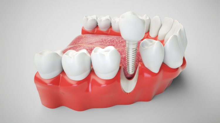
A dental implant restoration is the custom-made prosthetic component attached to an implant after the implant has integrated with the jawbone. Although patients often use the word “implant” to describe the entire replacement tooth, the implant itself is the supporting fixture. The restoration may be a crown, bridge, fixed full-arch prosthesis, or implant-retained overdenture, depending on the number of missing teeth and the patient’s clinical needs.
In my practice, I view implant restoration as a coordinated process involving biology, biomechanics, material science, and precise digital or conventional planning. The restoration must reproduce natural appearance and function while protecting the implant connection, surrounding bone, and peri-implant tissues. Its success depends on accurate fit, controlled occlusion, appropriate contours, cleansability, and long-term maintenance.
What a Dental Implant Restoration Actually Is
The Implant and the Restoration Are Different Components
When I discuss implant treatment with patients, I distinguish carefully between the dental implant and the dental implant restoration. The implant is the component positioned within the jawbone to function as an artificial tooth root, while the restoration is the prosthetic structure that recreates the visible and functional portion of the missing tooth. Depending on the clinical situation, that restoration may be a single crown, a multi-unit bridge, a fixed complete-arch prosthesis, or an implant-retained removable overdenture. These components function together, but they are designed, placed, evaluated, and maintained according to different biological and mechanical principles. An implant can be well integrated in bone while the restoration above it develops a separate technical problem, such as ceramic chipping, screw loosening, loss of retention, or unfavorable occlusal contact. Understanding that distinction allows us to diagnose complications accurately rather than referring to every problem simply as an implant failure.
A Restoration Is a Complete Prosthetic System
A dental implant restoration is better understood as an engineered prosthetic system than as a conventional crown placed on an artificial root. It includes the restorative material, the implant-abutment connection, the abutment or titanium base, the retaining screw or cement interface, the emergence profile, the proximal contacts, and the occlusal anatomy. Every one of these elements influences how the restoration transfers functional forces and interacts with the surrounding soft tissue. The final appearance is important, particularly in the anterior region, but appearance alone is not an adequate measure of success. A clinically successful restoration must also be cleansable, retrievable when appropriate, mechanically stable, compatible with the implant system, and harmonious with the patient’s bite. I therefore design the restoration as part of a biological and biomechanical environment rather than treating it as an isolated replacement tooth.
Why the Definition Matters Clinically
The distinction between implant placement and implant restoration has practical consequences throughout treatment. Surgical success establishes a stable foundation, but the prosthetic design determines how that foundation will be loaded during chewing, swallowing, clenching, and parafunctional activity. A poorly contoured restoration can make plaque removal difficult even when the implant itself is ideally integrated. An unfavorable screw-access position can compromise esthetics, occlusal anatomy, or retrievability, while an excessive cantilever can increase mechanical leverage on the implant complex. Conversely, a carefully designed restoration can help preserve tissue form, distribute force predictably, and simplify maintenance over many years. This is why contemporary implant dentistry should be planned from the desired final restoration backward to the surgical position rather than placing the implant first and attempting to solve the prosthetic limitations afterward.
How the Implant, Abutment, and Restoration Work Together
The Osseointegrated Implant Fixture
The implant fixture is usually a threaded titanium or titanium-alloy component placed into prepared bone, although other implant materials and designs exist. Its purpose is not merely to occupy the space previously filled by a root but to provide a stable interface capable of supporting a prosthesis under repeated functional loading. During healing, bone forms a direct structural relationship with the implant surface through the process commonly described as osseointegration. The implant does not reproduce every anatomical or sensory feature of a natural tooth root, particularly because it lacks a periodontal ligament. As a result, the implant responds differently to lateral force, impact, and subtle occlusal discrepancies. The restoration must compensate for these differences through controlled geometry, appropriate materials, and a carefully adjusted occlusal scheme.
The Abutment as the Transitional Component
The abutment connects the implant platform to the visible restoration and is one of the most consequential components in the prosthetic design. It establishes the vertical and horizontal transition from the relatively narrow implant connection to the broader dimensions of a clinical crown. Stock abutments are manufactured in predetermined shapes, whereas custom abutments can be designed for a particular tissue depth, restorative margin, emergence contour, and implant angulation. In some designs, a separate crown is cemented onto the abutment, while in others the crown and abutment assembly are retained by a prosthetic screw. The abutment’s material, height, angulation, connection geometry, and surface position can influence retention, esthetics, tissue support, and accessibility for maintenance. I therefore select an abutment according to the entire restorative problem rather than choosing it only because it physically fits the implant.
The Restoration as the Functional Interface
The restoration is the portion that contacts food, adjacent teeth, opposing teeth, the tongue, and the cheeks. Its contours determine how effectively the patient can clean around the implant and how food moves through the interproximal and cervical spaces. Its occlusal anatomy determines where force is applied and whether that force is directed predominantly along the implant’s long axis or converted into potentially unfavorable bending moments. Because an implant lacks the cushioning and proprioceptive characteristics of a periodontal ligament, small discrepancies may be less readily perceived by the patient. Those discrepancies can nevertheless contribute to screw loosening, material wear, fracture, or overload of the surrounding prosthetic system. The restoration must therefore integrate esthetics, phonetics, cleansability, contact relationships, and force management within a single design.
Restoratively Driven Diagnosis and Treatment Planning
Beginning With the Intended Tooth Position
In advanced implant treatment, I begin by defining where the final tooth or prosthesis should be positioned before determining where the implant should be placed. This involves evaluating the incisal edge or occlusal plane, facial contour, tooth width, gingival architecture, restorative space, phonetics, lip support, and relationship to the opposing dentition. A diagnostic wax-up or digital tooth arrangement can reveal whether the proposed tooth position is compatible with the available bone and soft tissue. It can also identify situations in which orthodontic treatment, tissue augmentation, bone grafting, enamel recontouring, or modification of an adjacent restoration may improve the result. When implant placement is directed only by the volume of available bone, the implant may emerge in a position that complicates the restoration. Restoratively driven planning aligns the surgical foundation with the prosthetic endpoint from the beginning.
Integrating Three-Dimensional Anatomy
Cone-beam computed tomography can provide three-dimensional information about bone volume, ridge anatomy, neighboring roots, anatomical spaces, and the anticipated trajectory of implant placement. These data are most useful when they are interpreted in relation to the intended restorative design rather than considered in isolation. A site may contain enough bone to accommodate an implant but still offer an unfavorable restorative trajectory, inadequate facial tissue support, or insufficient distance from an adjacent structure. Digital planning can superimpose the proposed tooth position onto the radiographic anatomy, allowing the restorative and surgical objectives to be assessed together. In selected cases, a surgical guide can transfer this plan to the mouth, although guided surgery does not eliminate the need for clinical judgment or intraoperative verification. The objective is not simply accurate implant placement but placement that supports a maintainable, mechanically sound, and anatomically credible restoration.
Evaluating Restorative Space and Prosthetic Volume
The dimensions required for an implant restoration vary substantially according to the implant connection, abutment design, restorative material, framework, and opposing dentition. Limited interarch space may restrict the thickness of restorative materials or make it difficult to create adequate structural strength without compromising hygiene access. Excessive restorative space can also be problematic because it may require longer crowns, replacement of lost gingival volume, or a prosthesis that incorporates both tooth-colored and soft-tissue-colored materials. In a full-arch case, the available prosthetic envelope must accommodate the framework, veneering material, screw-access channels, cleansable intaglio surface, and sufficient bulk around the implant connections. I also evaluate smile dynamics and lip support because replacing only teeth may not compensate for advanced loss of the supporting ridge. These spatial relationships should be diagnosed before surgery because implant positions cannot be predictably modified after osseointegration.
Osseointegration, Stability, and the Timing of Restoration
Biological Healing Before Definitive Loading
After implant placement, the surrounding bone undergoes a coordinated healing and remodeling response. Initial mechanical stability is derived from the relationship between the implant and the prepared osteotomy, while subsequent biological stability develops as new bone forms at the implant surface. These processes overlap, and the appropriate restorative timeline depends on more than the number of weeks that have passed. Bone quality, implant design, insertion stability, grafting, systemic health, site anatomy, occlusal risk, and the type of planned prosthesis can all influence the loading decision. A definitive restoration should not be used to compensate for uncertain integration or inadequate primary stability. I regard timing as a biological and mechanical decision rather than a fixed calendar formula.
Immediate, Early, and Conventional Loading
Implant restorations may be delivered according to immediate, early, or conventional loading protocols, but these terms do not indicate that every implant is suitable for every timeline. Immediate provisionalization can be valuable in selected anterior and full-arch cases because it may preserve appearance, support soft-tissue contours, and reduce the period during which the patient is without a fixed tooth. However, immediate placement of a provisional restoration does not necessarily mean that the restoration should bear unrestricted functional contact. The implant’s stability, restorative design, number and distribution of implants, parafunctional risk, and ability to control occlusion must all be evaluated. Inappropriately loading an unstable implant can interfere with integration, while excessively cautious treatment may extend care without adding a meaningful biological advantage. The correct protocol is therefore selected case by case and should include clearly defined prosthetic restrictions during healing.
Transitioning From Provisional to Definitive Restoration
A provisional implant restoration can serve as a diagnostic instrument rather than merely a temporary cosmetic replacement. It allows me to evaluate tissue response, phonetics, contact position, esthetic proportions, cleansability, occlusal behavior, and the patient’s adaptation before fabricating the definitive prosthesis. In the anterior region, the provisional contour can be modified incrementally to guide the peri-implant mucosa toward a more natural emergence profile. In a full-arch case, a provisional prosthesis may also test tooth position, vertical dimension, lip support, speech, and access for hygiene. Information from this phase can be transferred to the laboratory or digital design environment instead of recreating the final restoration without clinical reference. A well-managed provisional stage can therefore reduce uncertainty and improve the predictability of the definitive result.
The Major Types of Dental Implant Restorations
Single-Implant Crowns
A single-implant crown replaces one missing tooth without preparing the adjacent teeth as retainers. Although the restoration may appear straightforward, it requires precise control of implant position, contact relationships, emergence contour, material thickness, and screw-access orientation. The mesial and distal contacts must be designed with an understanding that adjacent natural teeth may continue to migrate over time while the osseointegrated implant remains relatively stationary. The crown must also reproduce the cervical form of a natural tooth while remaining accessible for cleaning around the implant. In posterior sites, the design must tolerate substantial cyclic loading without creating excessive cantilever or thin restorative areas. In anterior sites, small differences in tissue level, translucency, implant angulation, or facial contour can become visually significant.
Implant-Supported Fixed Partial Dentures
An implant-supported fixed partial denture uses multiple implants to support connected replacement teeth. Connecting units can improve load distribution, but it also creates additional requirements for passive fit, framework accuracy, interimplant alignment, and hygiene access. The pontic areas must be shaped to support appearance and phonetics without compressing tissue or creating inaccessible plaque-retentive spaces. Implant distribution is often more important than the simple number of implants because an unfavorable span or cantilever can increase bending forces. When implants are not parallel, restorative components or angled screw channels may be needed to obtain an acceptable path of insertion and access position. The design should also permit repair or retrieval because a technical complication in one area may otherwise affect the entire connected prosthesis.
Full-Arch Fixed Prostheses and Overdentures
Full-arch implant rehabilitation may involve a fixed prosthesis, a removable overdenture retained by implants, or a design that combines implant support with removable access for maintenance. A fixed full-arch prosthesis can provide excellent stability and function, but it requires sufficient restorative space, appropriate implant distribution, a rigid and accurately fitting framework, and an intaglio contour the patient can clean. An overdenture can replace teeth and lost tissue volume while allowing the patient to remove the prosthesis for hygiene, repair, and replacement of retentive components. The appropriate choice depends on bone anatomy, lip support, dexterity, esthetic requirements, financial considerations, maintenance capacity, and the amount of tissue that must be replaced. A fixed prosthesis is not inherently superior merely because the patient cannot remove it. In many cases, a well-designed overdenture offers a more maintainable and anatomically appropriate solution than a fixed restoration with compromised contours.

Capturing the Implant Position With Conventional and Digital Workflows
Why Implant Impressions Require High Accuracy
An implant impression must record the three-dimensional position and rotational orientation of the implant connection with sufficient accuracy for the restoration to seat without distortion. Unlike a prepared natural tooth, the implant platform is a manufactured interface with very limited tolerance for discrepancies between the clinical position and the working model or digital file. A small error in a single crown may alter contact or screw seating, while accumulated errors across multiple implants can prevent passive framework fit. Conventional techniques use impression copings and elastomeric materials to transfer implant position to a physical cast. Digital techniques use scan bodies with known geometries that allow software to calculate the implant platform’s location and orientation. Neither approach is automatically accurate unless the components are completely seated, stable, compatible, and captured according to a controlled protocol.
Digital Implant Scanning
Digital implant impressions can improve patient comfort, facilitate communication with the laboratory, and integrate efficiently with computer-aided design and manufacturing. During scanning, a precisely manufactured scan body is attached to the implant or intermediate abutment and recorded along with the surrounding teeth and soft tissue. The software identifies the scan body and substitutes the corresponding implant-library geometry, creating a virtual representation of the restorative connection. Accuracy depends on scan-body design, visibility, complete seating, scanner performance, operator technique, span length, tissue movement, and the amount of recognizable anatomical information available for image stitching. Clinical research supports the use of digital impressions for many implant indications, although full-arch applications remain more demanding than short-span restorations. I therefore select the impression method according to the complexity of the case rather than assuming one workflow is universally superior.
Verification in Complex Restorations
When several implants support a connected prosthesis, verification becomes especially important because minor transfer errors can accumulate across the arch. A verification index or jig can provide an independent clinical check of whether the recorded implant positions correspond accurately to the intraoral positions. Framework try-ins, sequential screw tests, tactile evaluation, radiographs, and digital or mechanical measurements may also contribute to the assessment of fit. No single method identifies every form of discrepancy, particularly when interfaces are subgingival or implants are deeply placed. If a framework appears to require screw tightening to draw it into position, I do not regard that as passive seating. The prosthesis should seat accurately before definitive torque is applied, because tightening screws should secure a correctly fitting restoration rather than force a misfitting one into place.
Abutment Selection and the Emergence Profile
Stock, Custom, and Titanium-Base Abutments
Abutment selection is guided by implant depth, tissue thickness, implant angulation, restorative space, margin position, material requirements, and the intended retention method. A stock abutment may be appropriate when its dimensions correspond closely to the clinical situation and its restorative margin can be positioned accessibly. A custom abutment offers greater control over emergence form, support of the definitive restoration, path of insertion, and location of the restorative margin. Titanium-base designs combine a prefabricated metallic implant connection with a bonded ceramic or metal superstructure, allowing digital customization while preserving a precise titanium interface at the implant. The height and geometry of the bonding surface must be adequate for the restorative material and anticipated loading. The choice should therefore reflect mechanical and biological requirements rather than being made solely on esthetic preference.
Developing the Transmucosal Contour
The emergence profile is the contour through which the restoration transitions from the implant platform to the shape of the clinical crown. In natural dentition, this transition is influenced by root anatomy, connective tissue attachment, bone position, and gingival form, none of which is duplicated automatically by a cylindrical implant. A provisional restoration can be modified to guide tissue maturation and create appropriate support for the facial and interproximal mucosa. Excessive convexity may compress tissue and reduce access for cleaning, whereas an undercontoured restoration may fail to support the intended soft-tissue architecture. The contour must also respect the depth and facial-palatal position of the implant because the restoration cannot compensate indefinitely for severe three-dimensional placement errors. I aim to create the necessary tissue support with the least bulky contour compatible with esthetics and function.
Margin Position and Cleanability
The position of a restorative margin has direct implications for cement removal, tissue health, scanning, impression accuracy, and future maintenance. Deep subgingival margins may sometimes be unavoidable, particularly when implant depth or esthetic requirements dictate the location of the crown-abutment junction. However, placing a margin deeper than necessary can make residual cement difficult to identify and remove. A custom abutment can often bring the margin into a more accessible position while preserving the required emergence contour. The restoration should also provide sufficient space for floss, interdental brushes, or other devices selected for the individual case. I consider a restoration incomplete if it appears attractive but cannot be cleaned effectively by the patient or maintained predictably by the clinical team.
Screw-Retained Versus Cement-Retained Restorations
Screw-Retained Implant Restorations
A screw-retained restoration is secured directly to the implant or to an intermediate abutment with a prosthetic screw. Its principal clinical advantage is retrievability because the restoration can often be removed for inspection, repair, hygiene access, screw replacement, or modification. This design also avoids a subgingival cement interface, which can reduce one important source of biological complication. The access channel must emerge in a location compatible with strength, esthetics, and occlusal anatomy, however, and unfavorable implant angulation may place it on the facial surface or near a critical contact. Screw-retained restorations can experience technical complications such as screw loosening, access-channel wear, or fracture of restorative material around the channel. Precise seating, correct component selection, appropriate torque, and controlled occlusion are therefore essential to their long-term performance.
Cement-Retained Implant Restorations
A cement-retained restoration resembles a conventional crown in that it is luted onto an implant abutment. This approach can provide an uninterrupted occlusal surface and may help manage an implant trajectory that would place a screw-access channel in an unacceptable position. Retention depends on abutment height, taper, surface area, surface treatment, cement selection, and the fit of the crown. The major biological concern is residual cement, particularly when the margin is positioned deeply below the gingiva and cannot be visualized or instrumented completely. Excess cement may contribute to inflammation, suppuration, or progressive peri-implant tissue breakdown in susceptible situations. When I use a cement-retained design, I place particular emphasis on accessible margin position, controlled cement volume, complete cleanup, and radiographic or clinical verification where appropriate.
Selecting the Retention Method
Neither screw retention nor cement retention should be selected according to a rigid rule that ignores implant position and prosthetic design. I evaluate retrievability, implant angulation, esthetic risk, restorative material, available occlusal space, abutment dimensions, tissue depth, anticipated maintenance, and the consequences of future repair. Angled screw-channel systems may allow a screw-retained restoration even when the implant axis is not ideally aligned, although the permitted correction and mechanical performance depend on the system. Some hybrid designs are extraorally bonded to a titanium base and then inserted as a screw-retained assembly, combining a customized ceramic restoration with a retrievable connection. Each additional interface introduces its own manufacturing, bonding, and maintenance requirements. The preferred design is the one that minimizes biological and technical risk for the specific implant position rather than the one that is most convenient during a single appointment.
Choosing Restorative Materials
Monolithic Zirconia
Monolithic zirconia is frequently selected for posterior implant restorations because it can provide substantial strength with less dependence on a layered veneering ceramic. Its high rigidity can be advantageous where restorative space is limited or occlusal loading is considerable, but rigidity alone does not guarantee clinical success. Connector dimensions, crown thickness, surface finish, opposing materials, titanium-base geometry, occlusal adjustment, and polishing all affect performance. Aggressive chairside adjustment followed by inadequate polishing may create a rough surface that accelerates wear of the opposing dentition. Highly translucent zirconia formulations can improve appearance but may not have the same mechanical characteristics as more opaque, high-strength formulations. I select the zirconia category and restoration design according to location, thickness, esthetic demand, and force rather than treating all zirconia as a single material.
Lithium Disilicate and Layered Ceramics
Lithium disilicate can provide favorable translucency and optical integration in selected implant restorations, particularly when sufficient material thickness and a supportive substructure are available. Its use requires careful attention to the location of the screw-access channel, the dimensions of the titanium base, bonding protocol, restoration thickness, and magnitude of occlusal force. Layered zirconia and metal-ceramic restorations can provide customized color and surface characterization but introduce a veneering layer that may be susceptible to chipping under unfavorable loading. In highly esthetic areas, a customized ceramic abutment or ceramic superstructure may help control the color beneath thin peri-implant tissue. That esthetic advantage must be balanced against the mechanical requirements of the connection and the depth of the implant. Material selection should therefore be based on the entire assembly rather than on the crown material considered alone.
Material Selection as a Risk-Based Decision
The strongest available material is not automatically the most appropriate material for every patient. I consider restorative space, implant diameter, implant position, cantilever, parafunction, opposing dentition, esthetic expectations, access-channel position, repairability, and the number of connected units. A patient with heavy bruxism and a posterior implant may require a different material strategy from a patient receiving a single lateral-incisor restoration. Full-arch prostheses introduce additional issues such as framework rigidity, shock absorption, tooth wear, veneering repair, and replacement of lost gingival volume. Material interfaces are also important because ceramic bonded to a titanium base behaves differently from a one-piece metal abutment supporting a separately cemented crown. The objective is not to select a fashionable material but to construct a prosthetic system whose individual components function coherently.
Occlusal Design and Force Management
Why Implant Occlusion Is Different
A natural tooth is suspended within the periodontal ligament, which permits microscopic movement and provides sensory feedback during function. An osseointegrated implant is ankylosed functionally to the surrounding bone and does not reproduce that same ligament-mediated response. This does not mean an implant restoration should be kept entirely out of occlusion, but it does mean that contact intensity and force direction require deliberate control. High contacts may be difficult for the patient to identify and can produce repeated loading of screws, restorative materials, and the implant-bone complex. Lateral and cantilevered forces generally create greater bending moments than forces directed closer to the long axis of the implant. I therefore evaluate implant occlusion dynamically rather than relying only on a static mark produced while the patient closes once on articulating paper.
Shaping Contacts and Occlusal Anatomy
The occlusal surface should provide functional efficiency without creating unnecessary leverage. Depending on the clinical situation, this may involve moderating cusp inclination, narrowing the functional table, positioning contacts centrally, reducing nonessential eccentric contacts, or limiting cantilever length. These adjustments should not produce a visibly undersized or anatomically implausible tooth, but they should acknowledge the implant’s position and the available restorative support. Contact design must also be coordinated with the opposing arch because an implant crown against a natural tooth behaves differently from one opposing another implant-supported restoration. In full-arch rehabilitation, the number and distribution of implants, framework design, anterior guidance, posterior contacts, and opposing prosthesis must be considered as one system. Occlusal design is therefore a patient-specific engineering decision rather than a standardized pattern applied to every implant crown.
Bruxism and Protective Strategies
Bruxism does not automatically exclude implant treatment, but it changes the mechanical risk profile. Repetitive clenching or grinding can contribute to ceramic fracture, screw loosening, component wear, loss of retention, and accelerated deterioration of opposing teeth or prostheses. Signs such as severe tooth wear, fractured restorations, hypertrophic masticatory muscles, recurrent screw problems, or a history of breaking appliances should be incorporated into treatment planning. I may modify restorative material, implant distribution, cantilever, connector dimensions, occlusal anatomy, or maintenance frequency when parafunctional loading is a concern. A protective occlusal appliance can be valuable in selected cases, but it must fit accurately and be monitored because the dentition and restorations can change over time. Mechanical risk cannot be eliminated completely, yet it can often be managed through design, surveillance, and early intervention.
Delivering and Verifying the Definitive Restoration
Evaluating Fit Before Final Retention
At the delivery appointment, I first confirm that the restoration seats completely without being pulled into position by the retaining screw. Proximal contacts, internal fit, tissue pressure, emergence contour, and the implant-abutment interface must all be evaluated before final torque or cementation. A contact that is excessively tight can prevent complete seating even when the restoration appears clinically close to the correct position. In a multi-unit restoration, sequential screw testing can help determine whether the framework is passive or whether tightening one area lifts another area away from its interface. Periapical radiographs can assist in evaluating subgingival connections when the imaging angle displays the interface clearly. A restoration that does not seat predictably should be corrected or remade rather than forced into place.
Applying the Correct Screw Torque
The retaining screw creates clamping force at the implant-abutment connection, and its performance depends on controlled preload rather than simply making the screw feel tight. Manufacturer-specific torque recommendations should be followed because screw dimensions, coatings, materials, connection geometry, and component design vary among implant systems. Under-tightening may provide inadequate preload, while excessive torque can deform or damage the screw or related components. The torque instrument must be appropriate for the system and maintained according to the manufacturer’s instructions. After torqueing, the screw-access channel is typically protected and sealed with materials that permit future retrieval without contaminating or damaging the connection. Occlusion is then rechecked because complete seating may alter contacts that appeared acceptable during the preliminary try-in.
Establishing a Clinical Baseline
The delivery visit should create a baseline against which future changes can be compared. I document the restoration design, implant system, components, torque specifications, occlusal contacts, tissue condition, probing findings when appropriate, and radiographic bone levels. Photographs and digital scans may provide additional reference information for esthetic areas or extensive rehabilitations. The patient should be shown exactly how to clean the restoration using devices that fit the actual prosthetic contour. Instructions should identify symptoms that warrant evaluation, including mobility, a changed bite, recurrent food trapping, bleeding, swelling, unpleasant taste, fracture, or loosening of an access-channel restoration. Long-term maintenance is more effective when both the clinical team and the patient know what the restoration looked and functioned like at the time of delivery.
Maintenance, Complications, and Long-Term Performance
Maintaining the Peri-Implant Tissues
Implant restorations require lifelong professional assessment and daily plaque control. The maintenance interval should be individualized according to periodontal history, tissue condition, prosthesis design, oral hygiene, smoking exposure, systemic risk factors, previous peri-implant disease, and the patient’s ability to clean the restoration. Professional visits may include examination of tissue color and contour, bleeding or suppuration, probing measurements, plaque accumulation, occlusion, mobility, screw-access integrity, and radiographic bone levels when indicated. Instruments and techniques should be selected to remove deposits without unnecessarily damaging the restoration or implant components. Full-arch restorations require particular attention because their intaglio contours and access spaces may make early inflammation difficult for the patient to detect. Supportive maintenance is not optional after implant treatment; it is part of the treatment itself.
Biological and Technical Complications
Peri-implant mucositis involves inflammatory changes in the soft tissue surrounding an implant, while peri-implantitis includes inflammation accompanied by loss of supporting bone. Bleeding, suppuration, increasing probing depth, radiographic bone change, or altered tissue contour should be investigated rather than assumed to be normal around an implant. Technical complications may include screw loosening, screw fracture, loss of cement retention, ceramic chipping, framework fracture, wear, contact opening, or fracture of the access-channel restoration. A biological and a technical complication can occur simultaneously because a loose or poorly contoured restoration may impair cleaning or alter force distribution. Early identification generally allows a wider range of conservative corrective options than delayed diagnosis. Patients should therefore report changes promptly even when the implant itself does not feel painful.
Longevity Depends on More Than Implant Survival
Implant-supported restorations can achieve high survival rates, but survival does not mean that a restoration will remain entirely maintenance-free. A functioning implant may require replacement of a worn screw, repair of a chipped surface, adjustment of an open contact, modification of the bite, treatment of inflamed tissue, or fabrication of a new restoration after years of service. Changes in the adjacent teeth, gingival margin, facial tissues, medical status, dexterity, or opposing dentition can alter a result that was initially satisfactory. Retrievable designs and complete records can make future maintenance substantially more manageable. In my view, long-term success means preserving comfort, function, tissue health, structural stability, esthetics, and the ability to intervene when normal biological or mechanical changes occur. A dental implant restoration works best when it is treated not as a permanent object that can be ignored, but as a sophisticated prosthesis supported by an ongoing program of clinical care.
To Conclude
A dental implant restoration is not simply an artificial tooth placed over an implant. It is a precisely designed prosthetic system that must integrate with the implant fixture, surrounding tissues, opposing teeth, and the patient’s overall bite. Successful treatment depends on restoratively driven planning, accurate component selection, appropriate materials, controlled force distribution, and meticulous delivery.
At Dental Implant Partners in San Francisco, I also emphasize that long-term success requires regular professional evaluation and effective home care. With thoughtful design and ongoing maintenance, an implant restoration can provide stable function, natural esthetics, and predictable service for many years.

Restore Your Smile With Dental Implant Partners
At Dental Implant Partners, we understand that a successful dental implant restoration depends on precise planning, advanced prosthetic knowledge, and attentive long-term care. Our practice has been led by Dr. Belinda Gregory-Head for more than 25 years and now includes an experienced team of prosthodontists and general dentists. Together, we provide restorative treatment ranging from fillings and veneers to complex dental rehabilitations supported by implants.
We approach every treatment carefully and ethically, with recommendations based on each patient’s oral health, functional needs, and personal goals. When implants are not the most appropriate option, we also provide expertly designed dentures and other restorative solutions. Our hygienists, who were trained as dentists and have cared for our patients for many years, play an important role in maintaining healthy teeth, implants, and surrounding tissues.
We love restoring smiles, and we would be honored to help you explore the right solution for your dental needs. If you are considering an implant-supported crown, bridge, or full-arch restoration, you can learn more about our approach to dental implants in San Francisco and how we plan treatment around long-term function, esthetics, and maintainability. Contact Dental Implant Partners to schedule a consultation and find out whether dental implant treatment is appropriate for you.